Structure of Long bone
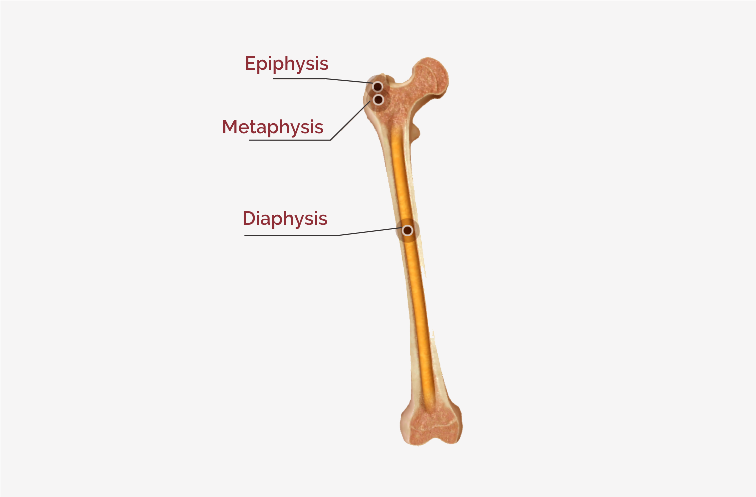
Here we see about the structure of long bone that has a greater length than width. The examples of long bone are-humerus, femur. A long bone consists of the following parts-
Diaphysis– It is the cylindrical shaft or body of the bone.
Epiphyses– These are the proximal and distal ends of the bone.
Metaphyses– These are the regions between the diaphysis and the epiphyses. In a growing bone contains an epiphyseal plate, a layer of hyaline cartilage that allows the diaphysis of the bone to grow in length. When a bone stops growing in length, the cartilage in the epiphyseal plate is replaced by bone, this structure is known as the epiphyseal line.
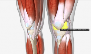
Articular cartilage- It is a thin layer of hyaline cartilage that covers the part of the epiphysis where the bone forms joint with another bone. It reduces friction and absorbs shock at freely movable joints.
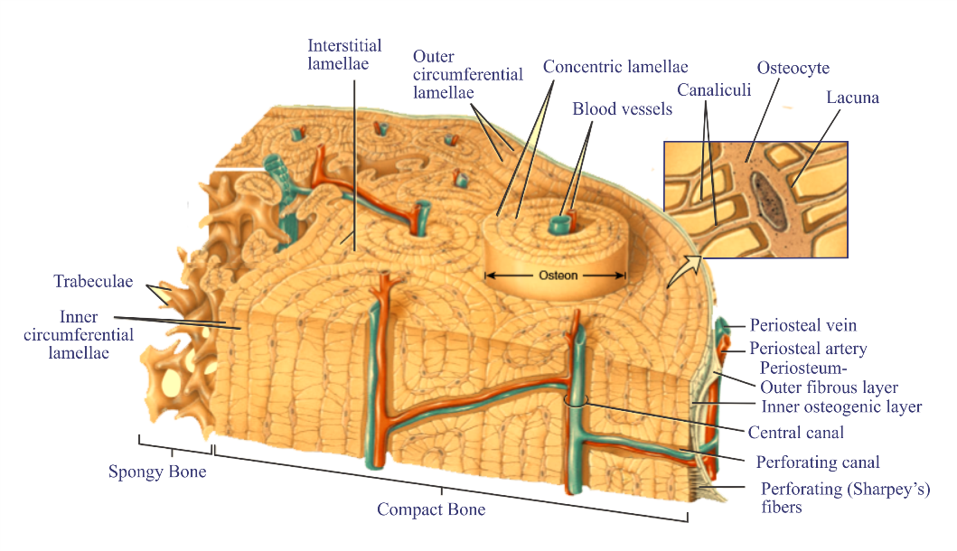
Periosteum– It is a connective tissue sheath associated with the blood supply that surrounds the bone surface wherever it is not covered by articular cartilage. It is composed of an outer fibrous layer of dense irregular connective tissue and an inner osteogenic layer that consists of cells it also protects the bone, nourishes bone tissue, and serves as an attachment point for ligaments and tendons. The periosteum is attached to the underlying bone by perforating fibers or Sharpey’s fibers, thick bundles of collagen that extend from the periosteum into the bone extracellular matrix.
The medullary cavity or marrow cavity– It is a hollow, cylindrical space within the diaphysis that contains fatty yellow bone marrow and numerous blood vessels. This cavity minimizes the weight of the bone by reducing the dense bony material.
Endosteum– It is a thin membrane that lines the medullary cavity. It contains a single layer of bone-forming cells and a small amount of connective tissue.
Blood supply-
-Bone is richly supplied with the blood.
-Periosteal arteries are small arteries, that enter the diaphysis through many perforating (Volkmann’s) canals and supply the periosteum and outer part of the compact bone.
-The center of the diaphysis, a large nutrient artery passes through a hole in the compact bone called the nutrient foramen. After entering the medullary cavity, the nutrient artery divides into proximal and distal branches that supply towards the end of the bone.
-These branches supply the inner part of compact bone tissue of the diaphysis and the spongy bone tissue and red bone marrow as far as the epiphyseal plates.
– The ends of long bones are supplied by the metaphyseal and epiphyseal arteries.
Veins that carry blood away from long bones are-
-One or two nutrient veins exit through the diaphysis.
-Epiphyseal veins and metaphyseal veins exit through the epiphyses and metaphyses.
-Small periosteal veins exit through the periosteum.