Layers of the Gastrointestinal Tract
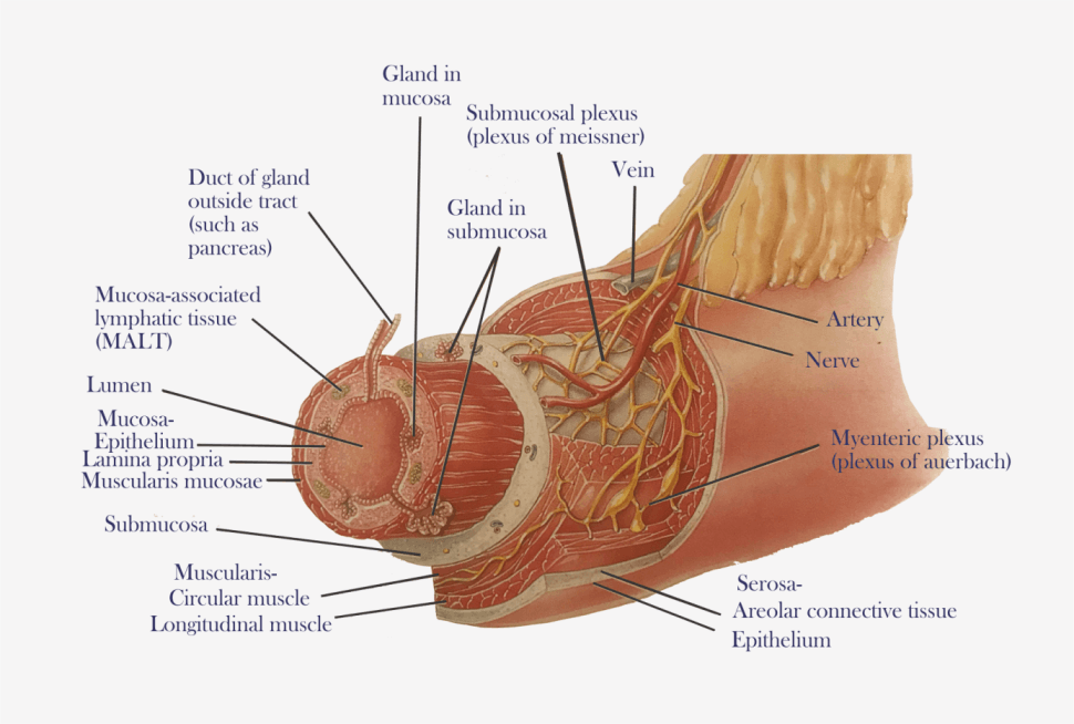
The wall of the GI tract from the esophagus to the anal canal has four-layer from deep to superficial, are the mucosa, submucosa, muscularis and serosa/adventitia.
A) Mucosa-
The mucosa, or innermost of the GI tract, is a mucous membrane. It is composed of epithelium, connective tissue (lamina propria) and a layer of smooth muscle (muscularis mucosa).
1. The epithelium in the mouth, pharynx, esophagus, and anal canal is stratified squamous epithelium that serves a protective function. Simple columnar epithelium, which functions in secretion and absorption, lines the stomach and intestines or firmly seal neighboring simple columnar epithelial cells to restrict leakage between the cells. Among the epithelial cells are few exocrine cells that secrete mucus into the lumen of the tract, and several types of endocrine cells, collectively called enteroendocrine cells, which secrete hormones.
2. The lamina propria is a areolar connective tissue containing many blood and lymphatic vessels, by which nutrients absorbed into the GI tract. This layer supports the epithelium and binds it to the muscularis mucosae. This contains the mucosa-associated lymphatic tissue (MALT), immune system cells that protect against disease.
3. Muscularis mucosae throw the mucous membrane of the stomach and small intestine into many small folds, which increase the surface area for digestion and absorption.
B) Submucosa-
The submucosa consists of areolar connective tissue that binds the mucosa to the muscularis. It contains many blood and lymphatic vessels that receive absorbed food molecules. The network of neurons in this layer known as the submucosal plexus.
C) Muscularis-
The muscularis of the mouth, pharynx, contains skeletal muscle that produces voluntary swallowing. It also forms the external anal sphincter, which permits voluntary control of defecation. The rest of the tract, the muscularis consists of smooth muscle with circular fibers inner and an outer sheet of longitudinal fibers. Contractions of the smooth muscle help break down food, mix it with digestive secretions, and propel it along the tract. Between the layers of the muscularis is a plexus of neurons the myenteric plexus.
D) Serosa-
A superficial layer called the serosa. It is a serous membrane composed of areolar connective tissue and simple squamous epithelium (mesothelium). The serosa is also called the visceral peritoneum because it forms a portion of the peritoneum. The esophagus lacks a serosa, only a single layer of areolar connective tissue called the adventitia forms the superficial layer of this organ.
Neural supply of the GI Tract-
1) Enteric Nervous System- the “brain of the gut,” consists of about 100 million neurons that extend from the esophagus to the anus. The neurons of the ENS are arranged into two plexuses: the myenteric plexus and submucosal plexus.
-The myenteric plexus or plexus of Auerbach is located between the longitudinal and circular smooth muscle layers of the muscularis.
– The submucosal plexus, or plexus of Meissner, is found within the submucosa.
-The plexuses of the ENS consist of motor neurons, interneurons, and sensory neurons. The motor neurons of the myenteric plexus supply the longitudinal and circular smooth muscle layers of the muscularis, which controls GI tract motility.
-The motor neurons of the submucosal plexus supply the secretory cells of the epithelium, controlling the secretions of the GI tract.
-The interneurons of the ENS interconnect the neurons of the myenteric and submucosal plexuses.
-The sensory neurons of the ENS supply the epithelium and contain receptors in the lumen of the GI tract like chemoreceptors, which respond to certain chemicals in the food present in the lumen, mechanoreceptors, as stretch receptors, that are activated when food stretches the wall of a GI organ.
2) Autonomic Nervous System-
– ENS is regulated by the neurons of the autonomic nervous system.
– The vagus (X) nerves supply parasympathetic fibers to most parts of the GI tract, the large intestine, which is supplied with parasympathetic fibers from the sacral spinal cord.
– The parasympathetic nerves that supply the GI tract form neural connections with the ENS. Parasympathetic preganglionic neurons of the vagus or pelvic splanchnic nerves synapse with parasympathetic postganglionic neurons located in the myenteric and submucosal plexuses.
-Stimulation of the parasympathetic nerves that supply the GI tract causes an increase in GI secretion and motility by increasing the activity of ENS neurons.
-Sympathetic nerves that supply the GI tract arise from the thoracic and upper lumbar regions of the spinal cord. The sympathetic nerves that supply the GI tract cause a decrease in GI secretion and motility by inhibiting the neurons of the ENS. Emotions such as anger, fear, and anxiety may slow digestion because they stimulate the sympathetic nerves that supply the GI tract.