Vertebral Column
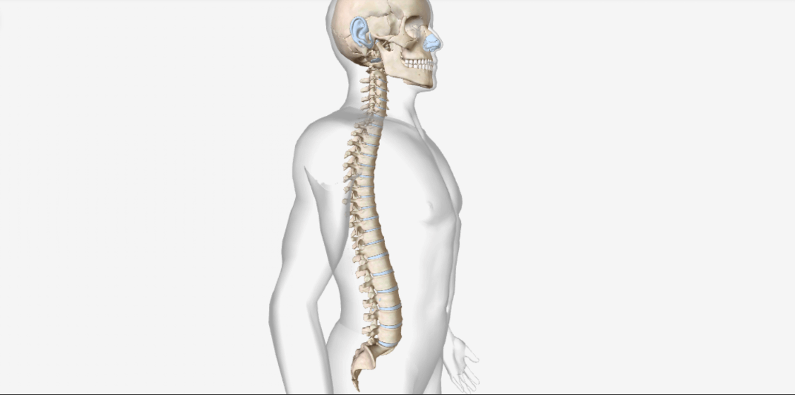
The vertebral column, also called the backbone, spinal column, or spine is composed of a series of bones called vertebrae.
-The vertebral column forms the part of the axial skeletal system. It consists of bone and the spinal cord and is about 71 cm long in males and 61 cm in the female.
-It forms a strong, flexible rod that can move forward, backward, and sideways, and rotate & supports the head and provide a point for attachment of the ribs, pelvic girdle, and muscles.
-It is not straight, rather it forms curves, the cervical, and lumbar curves are convex the thoracic and sacral curves are concave, as these increases the strength, absorb shock & maintain its position.
-The total number of vertebrae during early development is 33. As a child grows, the sacral and coccygeal vertebra fuses & contains 26 vertebrae- 7 cervical vertebrae in the neck region, 12 thoracic vertebrae, posterior to the thorax, 5 lumbar vertebrae supporting the lower back, 1 sacrum and 1 coccyx.
-There are intervertebral Discs between the bodies of adjacent vertebrae from the second cervical vertebra to the sacrum. Each consisting of an outer ring of fibrocartilage called the annulus fibrosus, and an inner elastic substance called the nucleus pulposus. The superior and inferior is a thin layer of hyaline cartilage.
-The parts of a typical Vertebrae in different regions of the spinal column vary in size, shape, a vertebral body, a vertebral arch, and several processes.
-The vertebral body is the thick, disc-shaped anterior portion, its superior and inferior surfaces are roughened, the anterior and lateral surfaces contain nutrient foramina, openings through which blood vessels enter.
-The 2 short, thick processes, the pedicles project posteriorly from the vertebral body and then unite with the flat laminae to form the vertebral arch.
-The vertebral body and the vertebral arch forms the vertebral foramen & contains the spinal cord, adipose tissue, areolar connective tissue, and blood vessels. All the vertebral foramina of all vertebrae form the vertebral canal or spinal canal.
-The pedicles have vertebral notches, they form an opening between adjoining vertebrae called intervertebral foramen, which is the passage of a spinal nerve.
-Seven processes arise from the vertebral arch. At the point where a lamina and pedicle join, a transverse process extends laterally on each side. A single spinous process projects posteriorly from the junction of the laminae. The four processes form joints with other vertebrae above or below. The two superior articular processes of a vertebra articulate with the two inferior articular processes of the vertebra immediately above them and the two inferior articular processes of that vertebra articulate with the two superior articular processes of the vertebra immediately below them. The articulating surfaces of the articular processes are the facets, covered with hyaline cartilage.
-The different vertebrae are as follows-
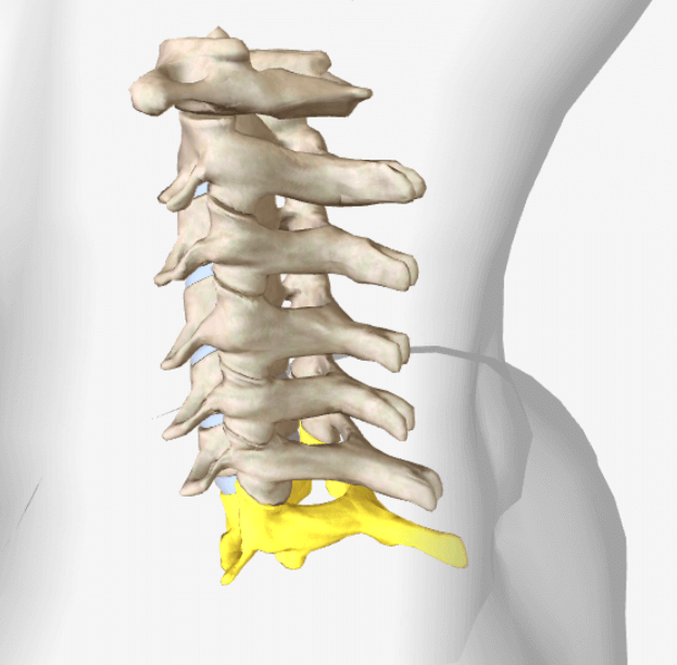
Cervical vertebrae (C1–C7)
-The bodies of the cervical vertebrae are smaller than all other vertebrae.
-Vertebral arches are larger.
-They have three foramina-one vertebral foramen and two transverse foramina, vertebral foramina are the largest as they have the cervical enlargement of the spinal cord & transverse process contains a transverse foramen through which the vertebral artery passes.
-The spinous processes of C2 through C6 are bifid as they have two projections at the tips.
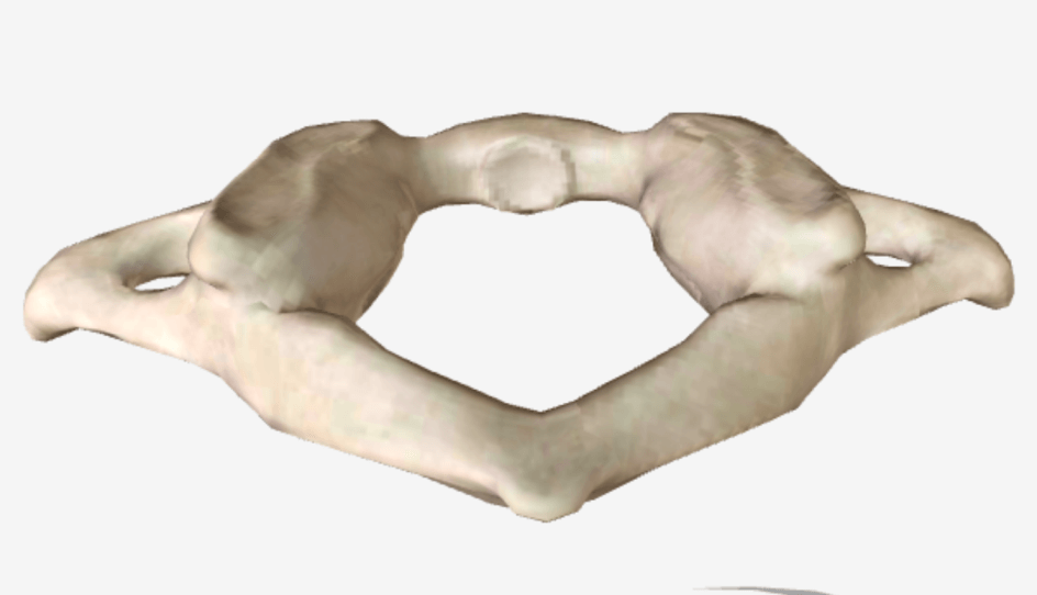
-The atlas (C1) is the first cervical vertebra is a ring-like bone with anterior and posterior arches and large lateral masses. It lacks a body and a spinous process. The superior articular facets, articulate with the occipital condyles of the occipital bone to form the paired atlanto-occipital joints.
-The inferior articular facets, articulate with the second cervical vertebra.
-In the second cervical vertebra (C2) the axis does have a vertebral body. A process called the dens or odontoid process projects through the anterior portion of the vertebral foramen of the atlas and makes a pivot on which the atlas and head rotate. This arrangement permits side to side movement of the head.
-The third through sixth cervical vertebrae (C3–C6), has the same structure as of a typical vertebra, seventh cervical vertebra (C7), called the vertebra prominens, has a large, nonbifid spinous process.
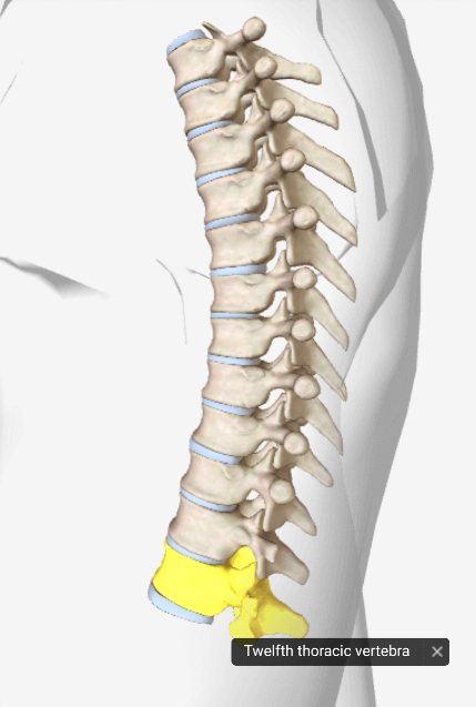
Thoracic vertebrae (T1–T12)
-These are larger and stronger than cervical vertebrae.
-The spinous processes of T1 to T10 are long and directed inferiorly and T11 and T12 are shorter, broader, and directed more posteriorly.
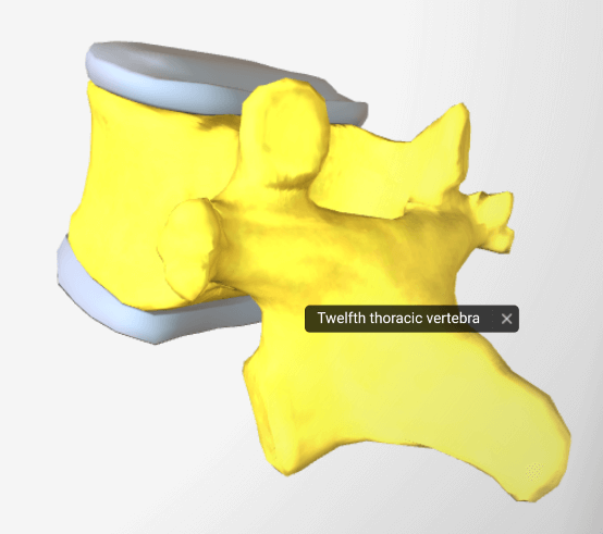
-These have longer and larger transverse processes with costal facets, the vertebral bodies of thoracic vertebrae have articular surfaces that form articulations with the heads of the ribs.
– A facet is formed when the head of a rib articulates with the body of one vertebra. Demifacet is formed when the head of a rib articulates with two adjacent vertebrae. These articulations between the thoracic vertebrae and ribs called vertebrocostal joints.

Lumbar vertebrae (L1–L5)
-These are the largest and strongest bones in the vertebral column.
-Their various projections are short and thick.
-The superior articular processes are directed medially and the inferior articular processes are directed laterally.
-The spinous processes are thick and broad, project posteriorly.
Sacrum (S1–S5)
-It is a triangular bone formed by the union of five sacral vertebrae.
-It serves as a strong pelvic girdle.

-The concave anterior side of the sacrum faces the pelvic cavity is smooth and contains four transverse lines that mark the joining of the sacral vertebral bodies.
-At the ends of these lines are four pairs of anterior sacral foramina. The lateral portion of the superior surface of the sacrum contains a smooth surface called the sacral ala.
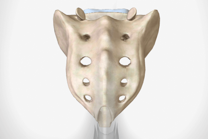
-The convex, posterior surface of the sacrum contains a median sacral crest, formed by the fused spinous processes of sacral vertebrae.
-The sacral canal is a continuation of the vertebral cavity.
-An inferior entrance to the vertebral canal called the sacral hiatus.
-On either side is a sacral cornu, which is connected by ligaments to the coccyx.
-The narrow inferior portion of the sacrum is known as the apex. The broad superior portion of the sacrum is called the base.
-Anterior projecting border of the base is the sacral promontory, the lateral surfaces the sacrum that articulates with the ilium of each hip bone to form the sacroiliac joint.
-The base of the sacrum articulates with the body of the fifth lumbar vertebra to form the lumbosacral joint.
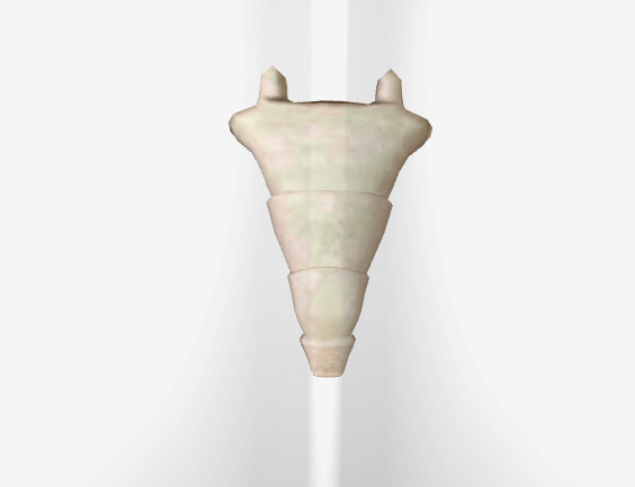
Coccyx (Co1–Co4)
-It is triangular in shape is formed by the fusion of four coccygeal vertebrae.
-The dorsal surface contains two long coccygeal cornua that are connected by ligaments to the sacral cornua.
-The coccygeal cornua are the pedicles and superior articular processes of the first coccygeal vertebra.
-The coccyx articulates superiorly with the apex of the sacrum.
-In females, the coccyx points inferiorly to allow the passage of a baby during birth.