Histology of bone
Here we see the microscopic structure of bones that contains an extracellular matrix that surrounds cells. The extracellular matrix consists of about 15% water, 30% collagen fibers, and 55% crystallized mineral salts like calcium phosphate these then combines with another mineral salt, calcium hydroxide, to form crystals.
As the crystals form, they combine with other mineral salts, like calcium carbonate and ions- magnesium, fluoride, potassium, and sulfate. As these mineral salts are deposited in the framework formed by the collagen fibers of the extracellular matrix, they crystallize and the tissue becomes hard. This process is called calcification.
There are four types of cells in bone tissue- osteogenic cells, osteoblasts, osteocytes, and osteoclasts.
-Bone stem cells, osteogenic cells derived from mesenchyme, & they undergo cell division or the resulting cells develop into osteoblasts.
–Osteoblasts are bone-building cells as they synthesize and secrete collagen fibers to build the extracellular matrix of bone tissue or they initiate the calcification process. As these osteoblasts surround themselves with the extracellular matrix, they become trapped in their own secretions and become osteocytes.
-Osteocytes are the mature bone cells, which maintain the daily metabolism, like the exchange of nutrients and wastes with the blood.
-Osteoclasts cells derived from the fusion of as many as 50 monocytes present in the endosteum. The side of the cell that faces the bone surface, is deeply folded into a ruffled border by its plasma membrane, where it releases lysosomal enzymes and acids that digest the protein and mineral components of the underlying extracellular bone matrix. This breakdown of the bone extracellular matrix is termed as resorption, it is a normal process.
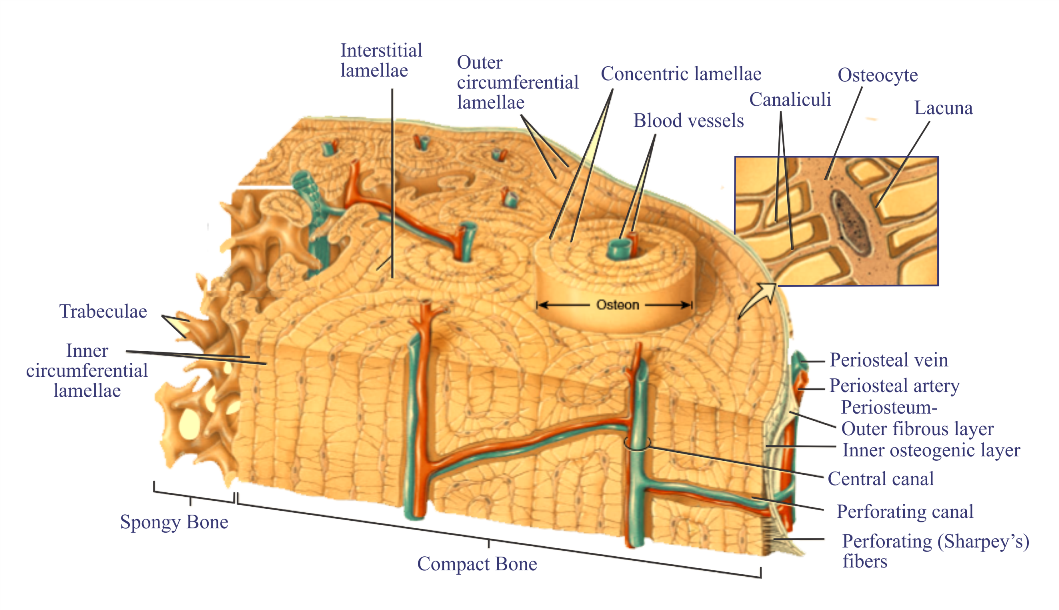
Depending on the size & distribution of the spaces, the regions of a bone may be categorized as compact or spongy, about 80% of the skeleton is compact bone and 20% is spongy bone.
Compact Bone Tissue
-It contains a few spaces and is the strongest form of bone tissue.
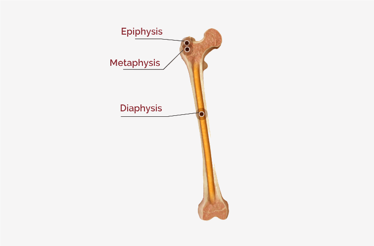
-It is present beneath the periosteum of all bones and forms the bulk of the diaphyses.
-It provides protection and support and resists the stresses produced by weight and movement.
-It is composed of repeating structural units called osteons, or Haversian systems.
-Each osteon consists of concentric lamellae arranged around a central canal or a Haversian canal.
-The concentric lamellae are circular plates of the mineralized extracellular matrix of increasing diameter or surrounds a network of blood vessels and nerves located in the central canal.
-These tube-like units form parallel cylinders that, runs parallel to the long axis of the bone.
-Between the concentric lamellae are small spaces called lacunae which contain osteocytes.
-Radiating in all directions from the lacunae are tiny canaliculi that are small channels, which are filled with extracellular fluid. Inside the canaliculi are fingerlike processes of osteocytes.
-The canaliculi connect lacunae with one another and with the central canals, forming an interconnected canal throughout the bone, so there are many routes for nutrients and oxygen to reach the osteocytes and for the removal of wastes.
-The areas between neighboring osteons contain lamellae called interstitial lamellae, which also have lacunae with osteocytes and canaliculi, these are fragments of older osteons that have been partially destroyed during bone rebuilding or growth.
-Blood vessels and nerves from the periosteum penetrate the compact bone through transverse perforating canals or Volkmann’s canals and connect with the medullary cavity, periosteum, and central canals.
-Arranged around the entire outer and inner circumference of the shaft of a long bone are lamellae called circumferential lamellae as they develop during initial bone formation.
-The circumferential lamellae deep to the periosteum are outer circumferential lamellae, connected to the periosteum by perforating fibers.
-The circumferential lamellae that line the medullary cavity are called inner circumferential lamellae.
Spongy Bone Tissue
-Also referred to as trabecular or cancellous bone tissue.
-It is located in the interior of a bone.
-It consists of lamellae that are arranged in an irregular pattern called trabeculae.
-Between the trabeculae are spaces that are filled with red bone marrow that produce blood cells, and yellow bone marrow & contains blood vessels that provide nourishment to the osteocytes.
-Each trabecula consists of concentric lamellae, osteocytes that lie in lacunae, and canaliculi that radiate outward from the lacunae.
-It is always covered by a layer of compact bone for protection.
-These are light thus reduces the weight of a bone so it allows the bone to move when it is pulled by a skeletal muscle.
-The trabeculae support and protect the red bone marrow.
-Spongy bone tissue in the hip bones, ribs, sternum, vertebrae, and the proximal ends of the humerus and femur is the only site where red bone marrow is stored and is the site of hemopoiesis that is blood cell production occurs in adults.