Introduction to Skeletal System
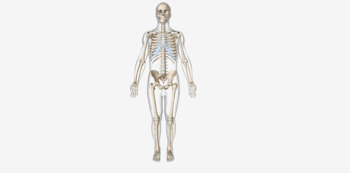
The skeletal system forms the framework of our body & there are 206 bones, paired (right and left sides of the body) or unpaired. The infants and children have more than 206 bones some of these then fuse later in life.
-There are 2 skeleton divisions: the Axial skeleton (80 bones) and the Appendicular skeleton (126 bones)
-The axial skeleton consisting of the bones that lie around the longitudinal axis of the body, a line that runs through the body’s center from the head to the space between the feet.
– The appendicular skeleton consists of the bones of the upper and lower limbs (extremities), or the bones forming the girdles that connect the limbs to the axial skeleton.
Let’s have a look into the table showing the name & numbers of bone that comes under the 2 divisions-
| Axial skeleton | Appendicular skeleton |
| –Skull includes cranium & face | –Pectoral/shoulder girdles include- |
| -Upper limbs include- | |
| –Thorax includes sternum & ribs | |
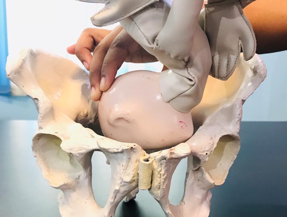
| – Pelvic/ hip girdle includes- | |
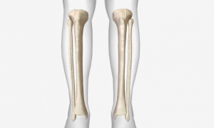
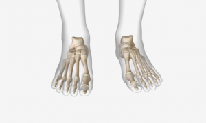
| -Lower limbs include- | |
| Total Number of bones = 80 | Total Number of bones = 126 |
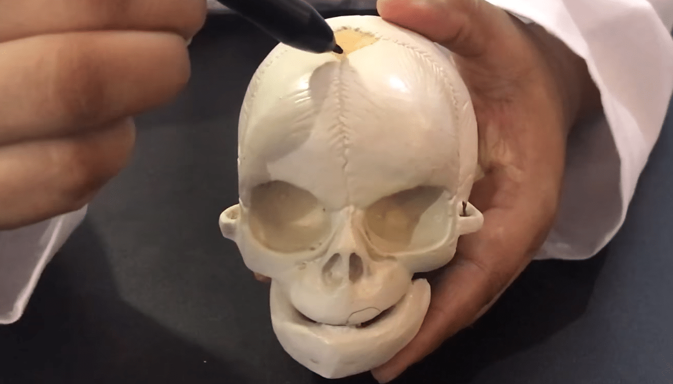
Types of Bones- On the basis of their shape, all bones are classified into five main types – long, short, flat, irregular, and sesamoid.
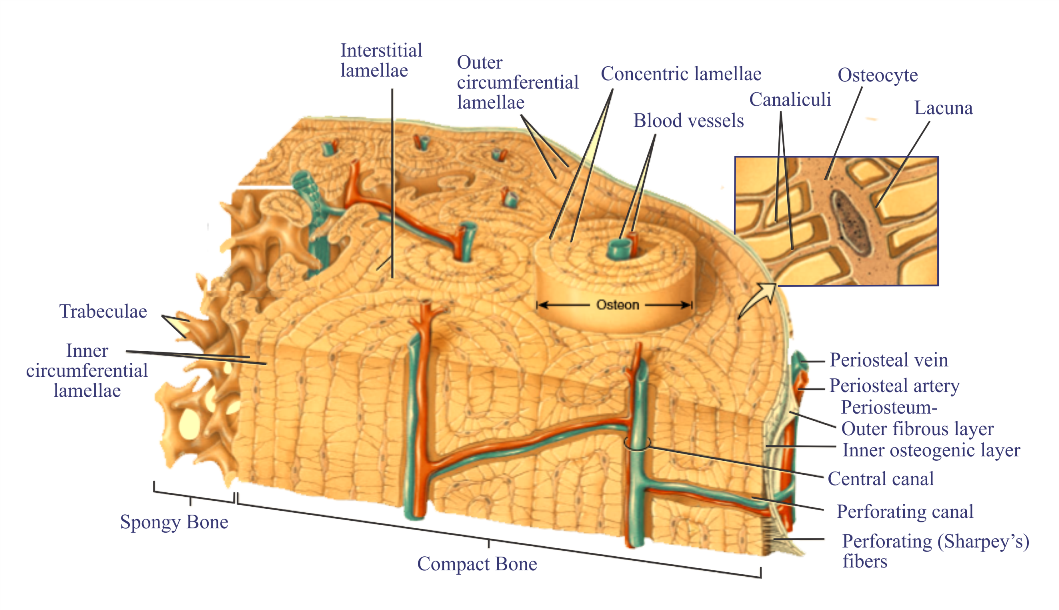
Long bones – It is mostly of compact bone tissue in their diaphyses & spongy bone tissue in their epiphyses ends, have a greater length than width, and are slightly curved to absorb the stress of the body’s weight at several different points. Examples- femur, tibia, fibula, humerus, ulna, radius and phalanges.
Short bones – These are of cube-shaped, there length and width are equal. They consist of spongy bone & on the surface a thin layer of compact bone tissue. Examples- carpal (wrist) bones and tarsal (ankle) bones.
Flat bones– These are thin and composed of two parallel plates of compact bone tissue enclosing a layer of spongy bone tissue, these provide a large area for muscle attachment. Examples- sternum, ribs, scapulae.
Irregular bones –These have no exact shape and differ in the amount of spongy and compact bone. Examples –vertebrae, hip bones, calcaneus, and few facial bones.
Sesamoid bones– These are shaped of a sesame seed. Examples – two patellae (kneecaps), these protect tendons from excessive tear.
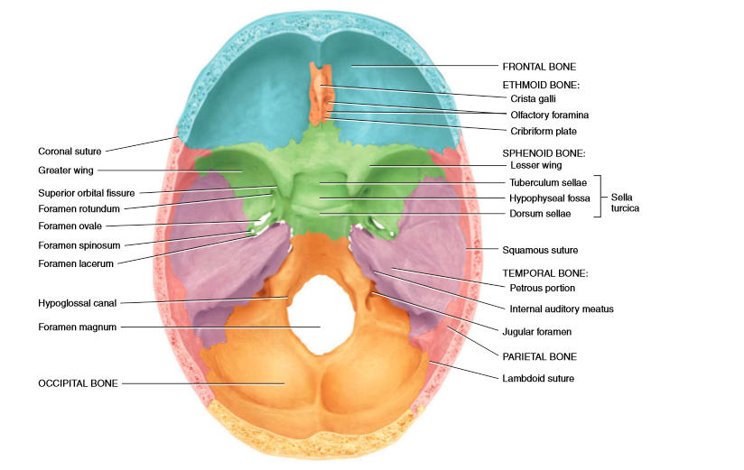
Bone Surface MarkingsThere are various structural adaptations on the bone for specific functions to be carried out as because of forces from tendons, ligaments. The below table will describe the various surface markings with the examples-
| (A) Depressions and openings- These are sites which allow the passage of soft tissue like nerves, blood vessels, ligaments, tendons or formation of joints, these as follows- | ||
| Fissure | The narrow slit between adjacent parts of bones through which blood vessels or nerves pass. | Superior orbital fissure of the sphenoid bone |
| Foramen | Opening through which blood vessels, nerves, or ligaments pass. | Optic foramen of the sphenoid bone |
| Fossa | Shallow depression | Iliac fossa of the hip bone |
| Sulcus | Furrow along the bone surface that accommodates blood vessel, nerve, or tendon. | coronary sulcus of heart |
| Meatus | Tubelike opening | External auditory meatus of the temporal bone |
| Processes- Projections or outgrowths on a bone that form joints or attachment points for ligaments and tendons, these as below- | ||
| Condyle | Large, round protuberance with a smooth articular surface at end of the bone | Lateral condyle of the femur |
| Facet | The smooth, flat, slightly concave or convex articular surface | The superior articular facet of vertebra |
| Head | Usually rounded articular projection supported on the neck (constricted portion) of bone | Head of the femur |
| Crest | Prominent ridge or elongated projection | Iliac crest of the hip bone |
| Epicondyle | The roughened projection above the condyle | Medial epicondyle of the femur |
| Line | Long, narrow ridge or border | Linea aspera of femur |
| Spinous process | Sharp, slender projection | The spinous process of vertebra |
| Trochanter | Very large projection | Greater trochanter of femur |
| Tubercle | Rounded projection | Greater tubercle of the humerus |
| Tuberosity | The variably sized projection that has a rough, bumpy surface | Ischial tuberosity of hip bone |
Functions–
Support– It provides a structural framework for the body by providing attachment points for the tendons of most skeletal muscles.
Protection– It protects the vital organs from injury. For example- cranial bones protect the brain, and the rib cage protects the heart and lungs.
Assist in movement– Skeletal muscles attached with the bones, they became contract & pull the bones to produce movement.
Mineral homeostasis– It stores calcium and phosphorus, which then strengthens the bone & also releases minerals into the bloodstream to maintain mineral balances & then distribute to other parts of the body.
Blood cell production– The bones contain red bone marrow which produces red blood cells, white blood cells, and platelets, this process is called hemopoiesis. It is present in developing bones of the fetus and in some adult bones, such as the hip bones, ribs, sternum, vertebrae, and skull. With increasing age, much of the bone marrow changes from red to yellow. Yellow bone marrow consists mainly of adipose cells, which store triglycerides.