Cranial bones
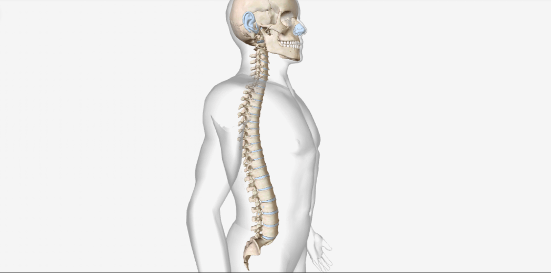
The skull is the part of the axial skeletal system which forms the bony framework of the head and rests on the superior end of the vertebral column, containing 22 bones of the cranium and the facial bones.
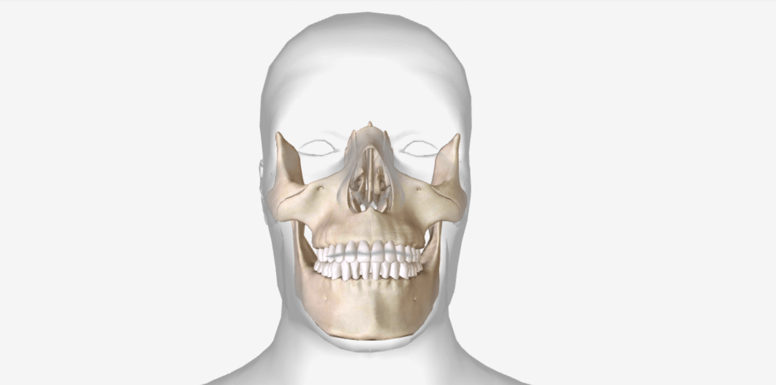
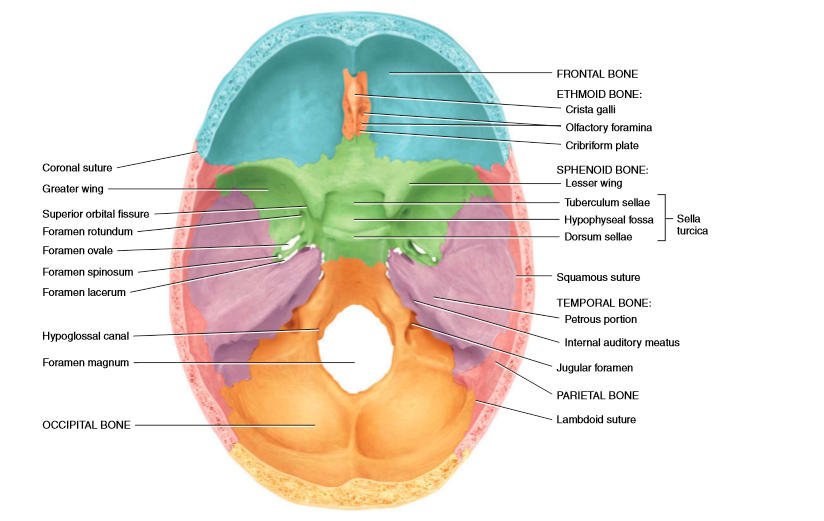
The total 8 cranial bones form the cranial cavity, which protects the brain, these are frontal bone, 2 parietal bones, 2 temporal bones, the occipital bone, the sphenoid bone, and the ethmoid bone.
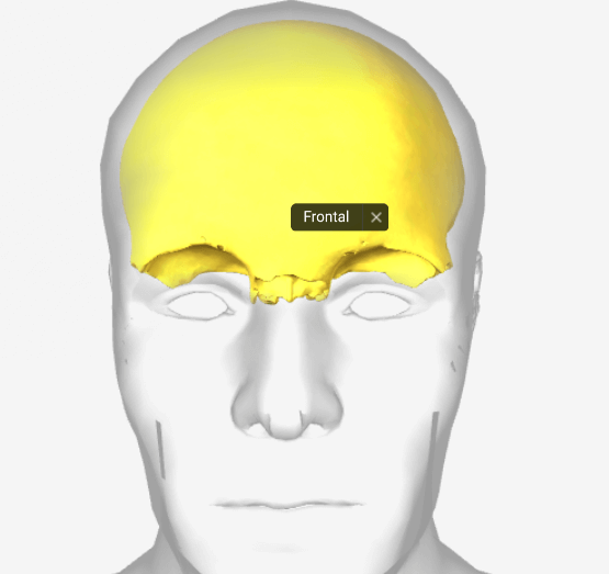
Frontal bone
-It forms the anterior part, the forehead, and the roof of the orbits.
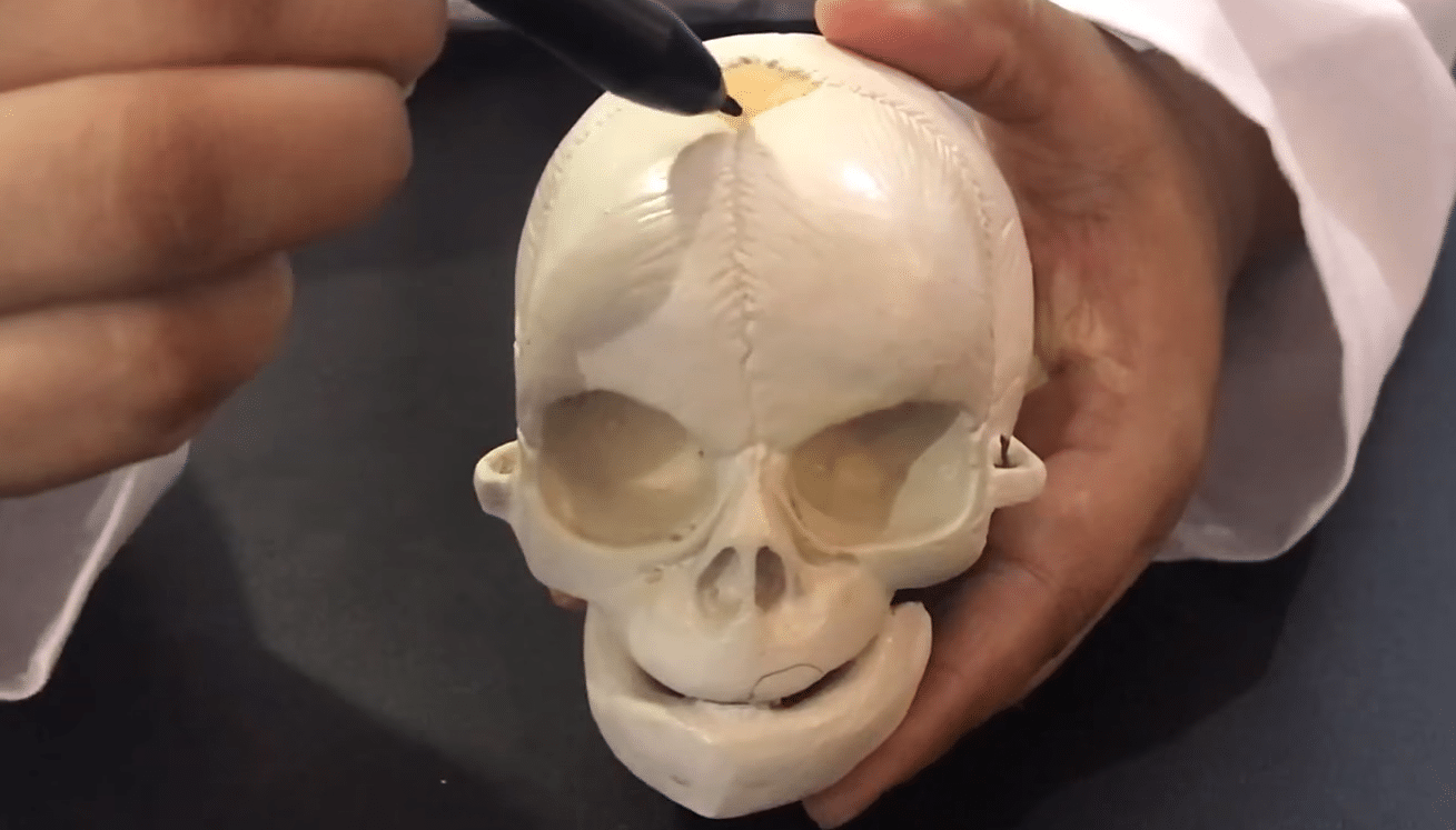
-After birth, the left and right sides of the frontal bone are united by the metopic suture, which disappears by the age of eight years.
-Frontal squama, a plate of bone that gradually slopes inferiorly from the coronal suture, becomes vertical above the orbits.
-At the superior border of the orbits, it thickens & forms the supraorbital margin.
-Within the supraorbital margin, at the midpoint, is a hole called the supraorbital foramen.
-The frontal sinuses lie deep to the frontal squama, are mucous membrane-lined cavities.

Parietal bones
–These paired bones form the sides and roof of the cranial cavity.
-The internal surfaces of the parietal bones contain many protrusions and depressions that accommodate the blood vessels supplying the dura mater, a covering of the brain.
Temporal bones
-These paired bones form the inferior lateral aspects of the cranium.
-The temporal squama a thin, flat part of the temporal bone forms the anterior and superior part of the temple.
-Projecting inferiorly from the temporal squama is the zygomatic process, which articulates with the temporal process of the zygomatic bone and forms the zygomatic arch.
-A socket called the mandibular fossa is located on the inferior posterior surface of the zygomatic process of each temporal bone. Anterior to the mandibular fossa is a rounded elevation, the articular tubercle.
-The mandibular fossa and articular tubercle articulates with the mandible to form the temporomandibular joint (TMJ).
-The mastoid portion is located posterior and inferior to the external auditory meatus which directs sound waves into the ear.
-The mastoid process is a rounded projection of the mastoid portion is the point of attachment for neck muscles.
-The internal auditory meatus is the opening through which the facial (VII) nerve and vestibulocochlear (VIII) nerve pass.
-The styloid process projects inferiorly and serves as a point of attachment for muscles and ligaments of the tongue and neck.
-At the floor of the cranial cavity a triangular part, located at the base of the skull between the sphenoid and occipital bones, contains the internal ear and the middle ear, helps in hearing and equilibrium.
-It also contains the carotid foramen, through which the carotid artery passes.
-Posterior to the carotid foramen is the jugular foramen, a passageway for the jugular vein.
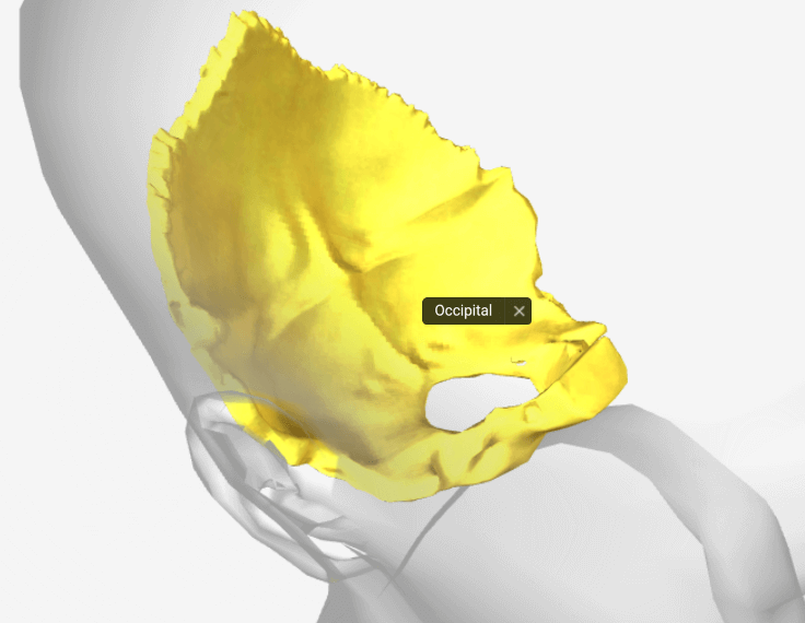
Occipital bone
-It forms the posterior part and the base of the cranium.
-The foramen magnum, a hole in the inferior part of the bone. The structure which passes through it is- the medulla oblongata that connects the brain with the spinal cord, the vertebral and spinal arteries or the accessory (XI) nerve.
-The occipital condyles, oval processes on either side of the foramen magnum, articulate with depressions on the first cervical vertebra the atlas to form the atlanto-occipital joint, which allows the head to move up & down.
-The external occipital protuberance, projection on the posterior surface above the foramen magnum through which ligamentum nuchae extends to the seventh cervical vertebra to support the head.
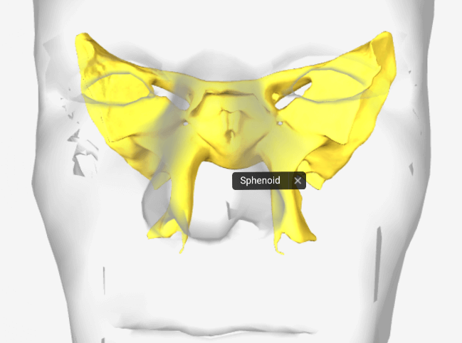
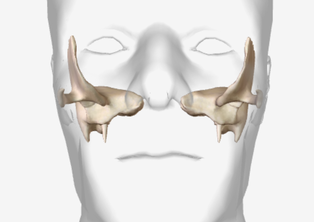
Sphenoid bone
-It lies at the base of the skull, forming a butterfly-shaped structure.
-It articulates with all the other cranial bones.
-It joins anteriorly with the frontal and ethmoid bones, laterally with the temporal bones, and posteriorly with the occipital bone.
-The sphenoid forms part of the floor, sidewalls, and the wall of the orbit.
-The body of the sphenoid is a hollow cube like portion between the ethmoid and occipital bones. The space inside the body is the sphenoidal sinus, which drains into the nasal cavity.
-The sella turcica is a saddle-shaped structure on the superior surface of the body of the sphenoid, anterior part forms the horn of the saddle, is tuberculum sellae, seat of the saddle is a depression, the hypophyseal fossa, which contains the pituitary gland and the posterior part forms the back of the saddle, is the dorsum sellae.
-The greater wings project laterally from the body and form the anterolateral floor of the cranium.
-The lesser wings are smaller, anterior and superior to the greater wings.
-Between the body and lesser wing is the optic foramen or canal through which the optic (II) nerve and ophthalmic artery pass into the orbit.
-The pterygoid processes project inferiorly from the points where the body and greater wings of the sphenoid bone unite some of the muscles that move the mandible attach to it.
-The foramen rotundum located at the junction of the anterior and medial parts of the sphenoid bone. The maxillary branch of the trigeminal (V) nerve passes through it.
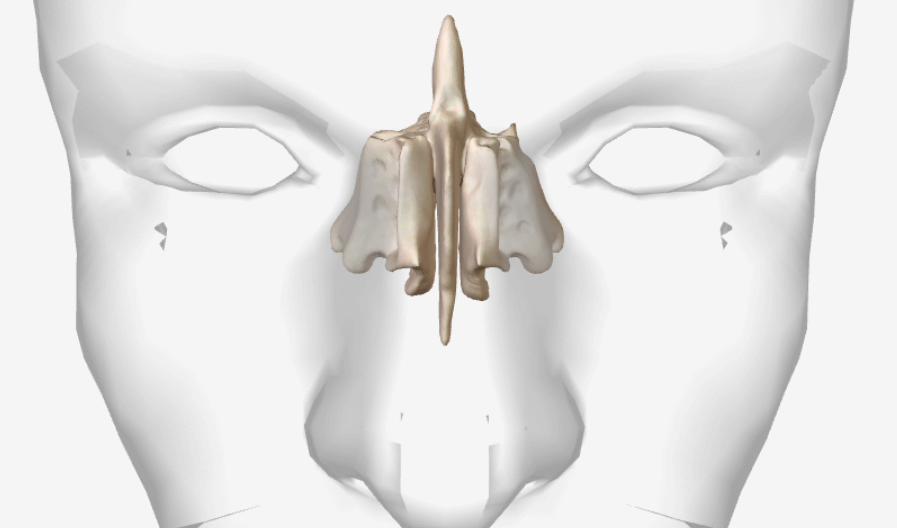
Ethmoid bone
-It is a sponge-like structure that forms the anterior part of the cranial floor medial to the orbits.
-It is a major superior supporting structure of the nasal cavity.
-The cribriform plate lies in the anterior floor of the cranium and forms the roof of the nasal cavity and contains the olfactory foramina through which the olfactory nerves pass.
-Projecting superiorly from the cribriform plate is a triangular process called the crista galli, it serves as a point of attachment for the falx cerebri, the membrane that separates the two sides of the brain.
-Projecting inferiorly from the cribriform plate is the perpendicular plate, that forms the superior portion of the nasal septum.
-The lateral masses of the ethmoid bone contains 3 to 18 air spaces called ethmoidal cells & together these cells form the ethmoidal sinuses.
-The lateral masses contain two projections lateral to the nasal septum, called as superior nasal concha and the middle nasal concha.
-The conchae increases the vascular and mucous membrane surface area in the nasal cavity, which warms and moistens inhaled air before it passes into the lungs.
-The conchae swirls the inhaled air so the particles become trapped in the mucus thus cleansing air before it passes into the lungs.