The placenta is a discoid shaped organ which is responsible for the exchange of waste and nutrient in between the mother and fetus.
Grossly, it has 2 surfaces- fetal and maternal or the peripheral margin, this occupies 30% of the uterine wall and 1/6th weight of the fetus at the term that is 500 gms. The fetal surface is smooth and glistening due to the presence of amnion in the inner aspect and the umbilical cord is attached to this surface at the center. The maternal surface is dull red, rough, and spongy and this surface is separated by the fissures.
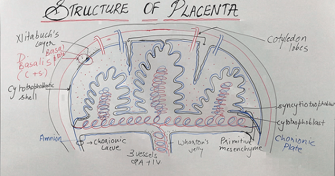
The internal structure consists of the 2 plates- the chorionic plate lies toward the fetus and the basal plate toward the mother and in between, two are the chorionic villi which are separated by decidua septum and form the functional unit of the placenta that is the cotyledon.
The chorionic plate is covered internally by the fetal membrane amnion and the layers within outward are- primitive mesenchyme containing the vessels, cytotrophoblast, and the syncytiotrophoblast. and the layers of the basal plate from outward to inward are- decidua basalis, Nitabuch’s layer cytotrophoblastic shell, and syncytiotrophoblast.
The umbilical cord contains 3 vessels 2 arteries and 1 vein and these covered by Wharton’s jelly.
Download the App: Android App
For more Lectures, please visit-
YouTube Channel – NursingLecture
Facebook – Facebook Page