Chorionic villi are the functional unit of the placenta which lies in between the basal plate or chorionic plate.
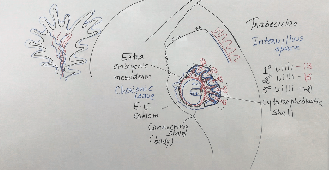
After the interstitial implantation, the outer syncytiotrophoblastic cells spread, and soon the spaces develop in between the cells that are called lacunae, and the syncytium cells erode the maternal capillaries too and drain maternal blood into the lacunae that eventually develops into the intervillous spaces. The syncytiotrophoblastic cells in finger-like projection and the lacunae in between them arrange in a cord-like fashion is called trabeculae.
Meanwhile, the embryoblastic cells form a bilaminar embryonic disc, one with the epiblastic layer forms an amniotic cavity and another with the hypoblastic layer forms the primary yolk sac. The yolk sac gives rise to the extra-embryonic mesoderm all around the embryoblast in between the embryoblast and cytotrophoblast. Soon few cells disintegrate to form a cavity within this mesoderm the extra-embryonic coelom.
On day 13 some of the cytotrophoblastic cells go into the finger-like projections of trabeculae and forms primary villi and when the mesodermal cell goes into these villi the called secondary villi by day 16 and these mesodermal cells start differentiating and forms blood vessels and cells within these finger-like projection called the tertiary villi by day 21. Meanwhile, some of the cytotrophoblastic cells in these villi penetrate the outer syncytiotrophoblastic cells and arrange them transversely by covering the syncytium layer forms the cytotrophoblastic shell.
The villi formation is taken place all around the blastocyst but the villi which are forming around the decidua capsularis becomes degenerated with the growing embryo and change to become the chorionic leave and forms the outer fetal membrane the is the chorion that is limited at the edge of the placenta. The epiblastic cells form the inner fetal membrane amnion.
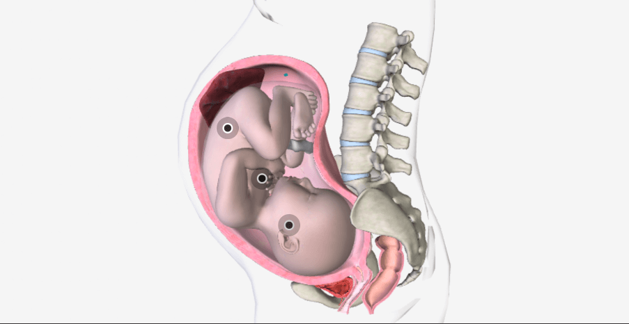
Few mesodermal cells attach with the caudal end of embryo to the cytotrophoblastic layer are not taking part in the formation of the coelom forms connecting band or body stalk and becomes the umbilical cord later on. This is attached to the center of the chorionic plate.
Download the App: Android App
For more Lectures, please visit-
YouTube Channel – NursingLecture
Facebook – Facebook Page