There are 4 Chambers in a Heart, 2 superior chambers the atrium are the receiving chambers and 2 inferior ventricles are the pumping chambers.
On the surface, there are 2 sulci- Coronary sulcus lies in between the paired atrium and the paired ventricles, and the anterior and posterior intraventricular sulcus lies between the ventricles anteriorly and posteriorly respectively.
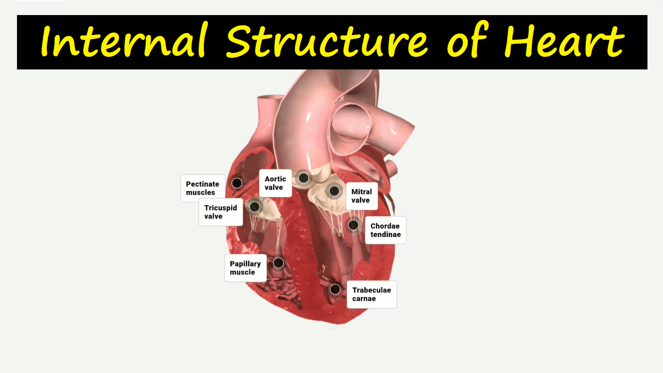
The right atrium receives blood through 3 vessels – Superior vena cava, inferior vena cava, and coronary sinus. Then with high-pressure blood from the right atrium moves into the right ventricle by a tricuspid valve (right atrioventicular or AV valve).
With a pressure rise in the right ventricle, blood ejects into the pulmonary trunk through the SL valve (pulmonary valve). In both ventricle the raised cardiac muscle bundles are called trabeculae carneae and some of the cone-shaped bundles are papillary muscles that attach with a tendon-like cord called chordae tendineae.
Blood in the left atrium is received by 4 pulmonary veins and with higher pressure, it moves into the left ventricle by a bicuspid valve (left atrioventricular valve or AV valve or Mitral valve).
From the rising pressure in the left ventricle, blood moves into the aorta by an aortic valve (SL valve), and as the ventricle relaxes blood returning back into the ventricle but free margins of the cusps closes the opening.
Download the App: Android App
For more Lectures, please visit-
YouTube Channel – NursingLecture
Facebook – Facebook Page