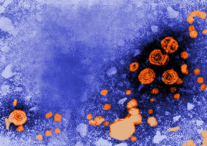
The infection of the liver caused by hepatotropic viruses is known as viral hepatitis.
There are five main types of hepatotropic viruses –
– Hep-A →RNA virus – feco-oral route
– Hep-B →DNA virus – parenterally transmitted
– Hep-C →RNA virus – post transfusion
– Hep-D →RNA virus –super infection with hepatitis B
– Hep-E →RNA virus –water-born contaminated (as after flooding)
(1.) Acute Hepatitis –
The most common consequence of all hepatotropic viruses is acute inflammatory involvement of the entire liver. In general type A,B,C,D,E show similar pathologic findings.
Grossly- The liver is slightly enlarged, soft & greenish.
Microscopically –
1) Hepatocellular injury – There is variation in the degree of liver cell injury.
-Mildly injured hepatocytes appear swollen with granular cytoplasm which ends to condense around the nucleus (ballooning degeneration)
– The nucleus becomes small & pyknotic & is eventually extruded from the cell, leaving behind necrotic mass called ‘councilman body’ by the process of apoptosis.
– Another type the hepatocellular necrosis is dropout necrosis in which isolated or small clusters of hepatocytes undergo lysis.
-Bridging necrosis is a more severe form of hepatocellular injury in acute viral hepatitis & may progress to chronic hepatitis.
2) Inflammatory infiltrates – Infiltration by mononuclear inflammatory cells, usually in the portal tract.
3) Kupffer Cell – Hyperplasia of kupffer cells contain phagocytosed cellular debris.
4) Cholestasis – Biliary stasis is not severe in viral hepatitis & may present as intracytoplasmic bile pigment granules.
5) Regeneration – Surviving adjacent hepatocytes undergo regeneration & hyperplasia.
(2.) Chronic Hepatitis –
It is defined as continuing or relapsing hepatic disease for more than 6 months with symptoms along with inflammation and necrosis. Pathologic features are common to both HBV, HCV infection.
- Piecemeal necrosis– It is defined as periportal destruction of hepatocytes at the limiting plate (piecemeal-piece by piece) expanded portal tracts are associated with proliferating bile ductules as a response to liver cell injury.
- Portal tract lesions-Inflammatory cell infiltration by lymphocyte, plasma cell, and macrophages. Proliferated bile ductules are expanded portal tracts.
- Intralobular lesions-there are the focal areas of necrosis and inflammation within the hepatic parenchyma, kupffer cell hyperplasia. A more severe form of injury shows bridging necrosis (bands of necrosed hepatocytes that are bridge portal tract to central vein)
- Bridging fibrosis– there is periportal fibrosis at the sites of interface hepatitis giving the portal tract stellate shaped appearance. End-stage of chronic hepatitis is characterized by dense collagenous septa destroying lobular architecture & forming nodules.