This article will discuss the bones, joints, and ligaments of the female pelvis.
Functions –
- The function of the pelvic girdle is to allow movement of the body, especially walking and running.
- It permits the person to sit or kneel.
- Women’s pelvis is adapted for childbearing and because of it’s increased width and rounded brim, women are less speedy than men.
- Pelvis transmits the weight of the trunk to the legs acting as the bridge between the femur.
- Pelvis takes the weight of the sitting body on the ischial tuberosity.
Pelvic bones –
There are 4 bones –
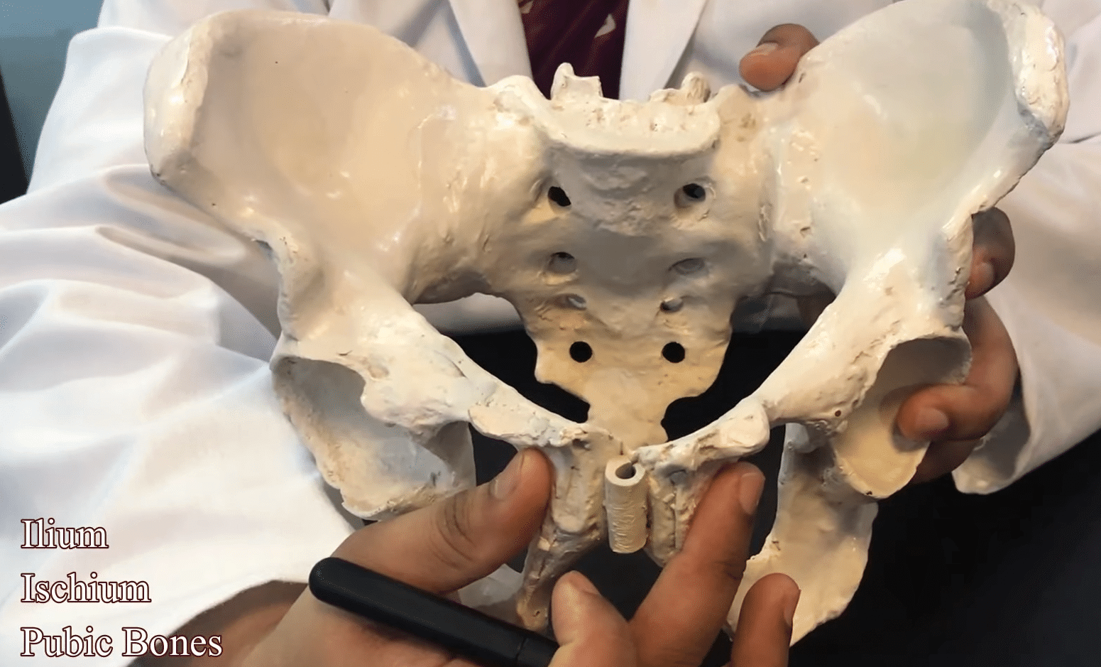
(1) INNOMINATE [NAMELESS] HIP BONE
Each bone have 3 parts –
ILIUM
It is the large flared-out part when the hand is placed on the hip it rests on the iliac crest, which is the upper border. At the front of the iliac crest, a bony prominence is known as the anterior superior iliac spine, a short distance below is the anterior inferior iliac spine, 2 similar points at the other end of the iliac crest, is posterior superior, posterior inferior iliac spine. The concave anterior surface of the ilium is an iliac fossa.
ISCHIUM
It is a thick lower part. It has large prominence known as ischial tuberosity on which the body rests when sitting behind & a little above the tuberosity is an inward projection, ischial spine.
PUBIC BONE
These bones form the anterior part. It has a body or 2 oars like projection- superior ramus & inferior ramus. The 2 public bones meet at symphysis pubis & 2 inferior rami form a pubic arch, the space enclosed by the body of the pubic bone, rami & ischium called obturator foramen.
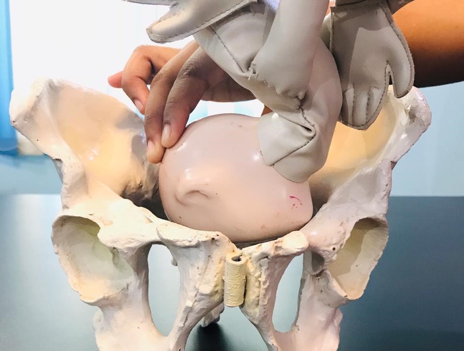
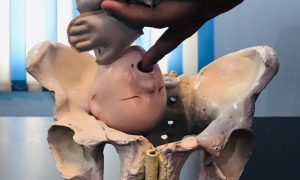
Hip bone contains a deep cut to receive the head of the femur. This is termed as acetabulum. On the lower border, 2 curves are formed. One extends from the posterior inferior iliac spine called the greater sciatic notch, the other lies between the ischial spine & ischial tuberosity are a lesser sciatic notch.
(2) THE SACRUM
It is a wedge-shaped bone consisting of five fused vertebrae. The upper border of the sacral vertebra just forwards is a sacral promontory. The anterior surface of the sacrum is concavely referred to as the hollow of the sacrum. Laterally extends into sacral ala or wing. 4 pairs of holes or foramina pierce the sacrum through these nerves from cauda equina emerges to pelvis organ. The posterior surface is rough to receive an attachment of muscles.
(3) THE COCCYX
It is a vestigial tail, consist of 4 fused vertebra, forming a small triangular bone.
Pelvic Joints-
There are 4 pelvic joints –
- One symphysis pubis – Is formed at the junction of 2 pubic bones, which are united by a pad of cartilage.
- Two sacroiliac joints – These are the strongest joints in the body. They join the sacrum to the ilium and connect the spine to the pelvis.
- Sacrococcygeal joint – This joint is formed where the base of the coccyx articulates with the tip of the sacrum.
In the non-pregnant state, there is very little movement in these joints but during pregnancy endocrinal activity causes the ligament to soften, which may provide more room for the fetal head as it passes through the pelvis.
Pelvic Ligaments-
Each of the pelvic joints is held together by a ligament.
- Interpubic ligament at the symphysis pubis
- Sacroiliac ligament
- Sacrococcygeal ligament
And the other 2 ligaments are –
- The Sacrotuberous ligament runs from the sacrum to the ischial tuberosity.
- Sacrospinous ligament from sacrum to the ischial spine.
Download the App: Android App
For more Lectures, please visit-
YouTube Channel – NursingLecture
Facebook – Facebook Page